In this tutorial, I use the Alphafold2 interface embedded in Chimera X to predict the structure of Cytochrome C from Skipjack Tuna. This tutorial teaches the student how to obtain the primary structure of the protein from the UniProt database, enter that into the Alphafold2 interface in Chimera X, and execute the code using Google Colab. After the structure is predicted, the experimental structure is downloaded from the Protein Data Bank (PDB) and superimposed with the predicted structure for comparison using Matchmaker in Chimera X.
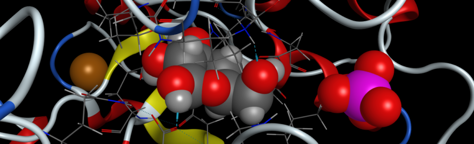
The protein, Cytochrome C (Cyt C) is a small (~100-150 residue) heme protein that is a part of the electron transport chain in mitochondria. This water-soluble protein is loosely associated with the inner mitochondrial membrane where it’s function is to transfer electrons from Coenzyme Q – Cytochrome C Reductase (Complex III) to Cytochrome C Oxidase (Complex IV). The electron transfer function is carried out by the oxidation and reduction of the iron atom at the center of the heme prosthetic group. It is interesting to note that Alphafold2 will predict the protein structure of Cytochrome C with great accuracy, yet it will not predict the structure of the heme prosthetic group. Yet that can be added with superimposition.
Procedure
- If you have not downloaded Chimera X, it can be obtained for Windows, OS X or Linux from the Resource for Biocomputing, Visualization, and Informatics (RBVI) at the University of California at San Francisco. The direct link to download the software is here.
- You will also need to obtain a free Google account to use Google Colab. While you can pay a small monthly fee to use Colab resources for priority use, it is not necessary to pay any fee for this tutorial.
- Locate the entry in the UniProt database for “Skipjack Tuna Cytochrome C“. It should have a UniProt Entry ID of P00025. Find the sequence in FASTA format and save that file to your computer, or cut-and-paste the sequence to your clipboard (Control-C).
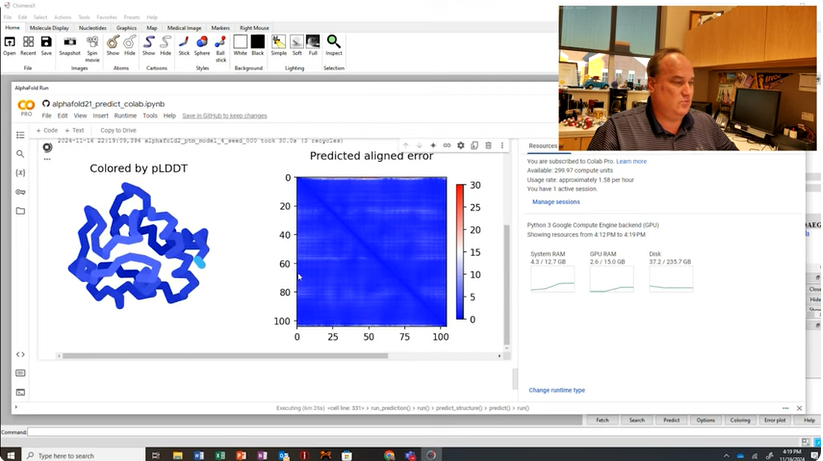
- Open Chimera X and go to TOOLS > STRUCTURE PREDICTION > ALPHAFOLD. To predict the structure using the sequence of Cytochrome C that you obtained from UniProt, copy the sequence to the Sequence window in the console on the right (Control-V) and click Predict. This will open a pop-up window for Google Colab where you will need to login with your Google username and password. Once logged in, execute the code and Google Colab’s servers should finish in approximately 10-20 minutes (assuming resources are currently available).
- Once the code finishes executing, it will sent the “best model” predicted to the visualization window in Chimera X. The model’s ribbons will be colored according to the confidence prediction, pLDDT score. The top five predicted models from Alphafold2 are also available and deposited in the Downloads/ChimeraX/AlphaFold directory (this directory may be located elsewhere depending on your specific installation of Chimera X).
- To compare the predicted structure with the experimental X-ray crystal structure, type “open 1CYC” in the Command: field at the bottom of Chimera X. Go to TOOLS > STRUCTURE ANALYSIS > MATCHMAKER to superimpose the two structures. Select 1CYC as the reference structure and the predicted structure and select the radio button for “Best-aligning pair of chains between reference and match structure“.
Questions
- Describe the key steps involved in predicting the structure of Cytochrome C using AlphaFold2 and visualizing it in ChimeraX. Why is it important to compare the predicted structure with the experimentally-determined structure?
- What type of sequence data is required for AlphaFold2 to predict the structure of Cytochrome C? How is this data prepared and formatted for use in the tutorial?
- What are the primary features of ChimeraX used in this tutorial for visualizing and analyzing the predicted structure? How do these features facilitate structural comparison?
- In the tutorial, how is the predicted structure of Cytochrome C superimposed onto the experimentally determined structure? What are the criteria used to assess the accuracy of the prediction?
- Why is the structure of Cytochrome C an appropriate choice for this exercise? Discuss its significance in biochemistry and how structural insights contribute to our understanding of its function.